Mestinon dosages: 60 mg
Mestinon packs: 30 pills, 60 pills, 90 pills, 120 pills, 180 pills, 270 pills
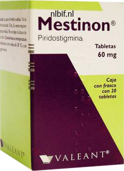
Purchase 60 mg mestinon otc
Aged muscle spasms 8 weeks pregnant buy discount mestinon 60mg on line, broken spasms small intestine cheap 60mg mestinon with visa, or abnormal pink blood cells are trapped by macrophages associated with uncommon vascular sinuses in the red pulp spasms head 60mg mestinon with amex. These macrophages break down the pink cells muscle relaxer kick in generic mestinon 60 mg with amex, start the metabolic breakdown of hemoglobin, and retrieve and retailer the iron from the heme for reutilization within the formation of latest pink blood cells in the bone marrow. The white pulp, however, is so named as a end result of its content of lymphocytes appears in life as whitish areas. In tissue sections, nevertheless, the nuclei of the closely packed lymphocytes impart an overall blue-staining response. The lymphatic tissue that constitutes the white pulp differs from nodules seen elsewhere in that it follows and ensheathes a blood vessel, the central artery. The lymphatic tissue surrounding the artery reveals periodic expansion, thus forming the nodules. This determine reveals, at the next magnification, the pink pulp and a portion of the trabecular vein from the world enclosed in the uppermost rectangle in the prime determine. In this specimen, the venous sinuses may be seen to advantage as a outcome of the purple blood cells in the sinuses have lysed and seem as unstained "ghosts"; only the nuclei of the white cells are readily seen. The wall of the vein is skinny, but the trabecula (T) containing the vessel offers the appearance of being part of the vessel wall. In people in addition to in different mammals, the capsule and the trabeculae that stretch from the capsule include myofibroblasts. Under situations of accelerating physical stress, contraction of these cells will happen and trigger speedy expulsion of blood from the venous sinuses into the trabecular veins and, thus, into the final circulation. This figure reveals, at larger magnification, the splenic nodule in the rectangle in the right portion of the figure above. Small arterial vessels and capillaries, branches of the central artery, provide the white pulp, and some pass into the reticular network of the marginal zone, terminating in a funnel-shaped orifice. Venous sinuses are also found in the marginal zone, and sometimes, arterial vessels could open into the sinuses. The details of the vascular provide are, at best, tough to resolve in typical H&E preparations. The penicillar arterioles, the terminal branches of the central artery, provide the red pulp but are likewise difficult to resolve. Thus, the relatively clear areas with scattered nuclei characterize the lumen of the venous sinus; the nuclei are these of white blood cells. In this specimen, the red blood cells have been lysed leaving only a transparent define Red pulp, spleen, human, H&E 1,200. This micrograph is a excessive magnification of the area within the rectangle of the previous micrograph. Other than the lysed red blood cells, which appear as empty circular profiles, a variety of lymphocytes (Ly) are current in the lumen. A slender however clearly seen intercellular house is present between adjoining cells. Also, processes of macrophages positioned outside of the sinuses in the splenic cords prolong between the endothelial cells and into the lumen of the sinuses to monitor the passing blood for overseas antigens. A macrophage (M), identified by residual bodies in its cytoplasm, is seen simply outdoors of the sinus. At the top of the micrograph, two venous sinuses (arrows) may be seen emptying into the trabecular vein. These small trabecular veins converge into larger veins, which ultimately unite giving rise to the splenic vein. The structural components which may be stained by the silver within the nodule encompass reticular fibers. The fantastic, thread-like stained materials that encircles the venous sinuses is a usual modification of basement membrane. Where the vessel has been cut deeper along its lengthy axis, the basement membrane appears as dot-like buildings (arrowheads). A threedimensional reconstruction of the basement membrane would reveal it as a sequence of ring-like constructions. The supporting reticular stroma arises from endodermal epithelium and produces a cellular reticulum.
Chilli (Capsicum). Mestinon.
- What other names is Capsicum known by?
- Are there safety concerns?
- What is Capsicum?
- Pain from shingles when applied to the skin.
- Dosing considerations for Capsicum.
- Are there any interactions with medications?
- Colic, cramps, toothache, blood clots, fever, nausea, high cholesterol, heart disease, stomach ulcers, heartburn, irritable bowel syndrome, migraine headache, allergic rhinitis, perennial rhinitis, nasal polyps, muscle spasms, laryngitis, swallowing dysfunction, and other conditions.
Source: http://www.rxlist.com/script/main/art.asp?articlekey=96908
Mestinon 60mg line
Classification of connective tissue is based on the composition and group of its extracellular components and on its features muscle relaxant drug list mestinon 60mg overnight delivery. One kind muscle relaxant starting with b best 60mg mestinon, the fibroblast muscle relaxant 1 order 60mg mestinon otc, produces the extracellular fibers that serve a structural function in the tissue spasms esophagus problems order mestinon 60 mg visa. In distinction, bone Connective tissue encompasses quite lots of tissues with differing functional properties but with sure common traits that allow them to be grouped together. Mesoderm, the center embryonic germ layer, gives rise to nearly all the connective tissues of the physique. The elastic fibers appear as blue-black, thin, lengthy, and branching threads without discernible beginnings or endings. Collagen fibers seem as orange-stained, lengthy, straight profiles, and are significantly thicker than the elastic fibers. Eosinophils and neutrophils (when present) may be identified by their distinctive segmented nuclei and presence of particular granules (reddish within the case of eosinophil). Note the different cell sorts most frequently found in the loose connective tissue with the surrounding extracellular matrix, which contains blood vessels and three different varieties of fibers. Maturation and proliferation of the mesenchyme give rise not only to the assorted connective tissues of the grownup but also to muscle, the vascular and urogenital techniques, and the serous membranes of the body cavities. The method during which the mesenchymal cells proliferate and arrange units the stage for the type of mature connective tissue that will kind at any particular website. Embryonic connective tissue is present in the embryo and inside the umbilical cord. The tissues normally cited as examples of elastic tissue are sure ligaments related to the spinal column and the tunica media of elastic arteries. The figuring out characteristic of reticular tissue is the presence of reticular fibers and reticular cells together forming a three-dimensional stroma. Reticular tissue serves as the stroma for hemopoietic tissue (specifically the red bone marrow) and lymphatic tissue organs (lymph nodes and spleen but not the thymus). Processes lengthen from these cells and get in touch with comparable processes of neighboring cells, forming a three-dimensional mobile community. The paucity of collagen fibers is according to the limited physical stress on the rising fetus. The spindle-shaped cells are broadly separated and seem much like fibroblasts within the near-term umbilical twine. The ground substance, nonetheless, is plentiful; in reality, it occupies more volume than the fibers do. It has a viscous to gel-like consistency and performs an necessary position within the diffusion of oxygen and vitamins from the small vessels that course via this connective tissue in addition to within the diffusion of carbon dioxide and metabolic wastes back to the vessels. Loose connective tissue is primarily situated beneath the epithelia that cover the body surfaces and line the internal surfaces of the physique. It can be related to the epithelium of glands and surrounds the smallest blood vessels (Plate 4, web page 188). This tissue is thus the preliminary site where pathogenic agents corresponding to micro organism that have breached an epithelial surface are challenged and destroyed by cells of the immune system. Most cell types in free connective tissue are transient wandering cells that migrate from local blood vessels in response to particular stimuli. Loose connective tissue is, due to this fact, the positioning of inflammatory and immune reactions. In areas of the physique where international substances are regularly current, giant populations of immune cells are maintained. For example, the lamina propria, the unfastened connective tissue of mucous membranes, corresponding to these of the respiratory and alimentary systems, contains giant numbers of those cells. Dense irregular connective tissue is characterised by ample fibers and few cells. Although morphologically the mesenchymal cells appear as a homogeneous population, they provide rise to cells that may differentiate into numerous cell varieties. The extracellular part of the tissue accommodates a sparse arrangement of reticular fibers and ample floor substance.
Proven 60mg mestinon
Dark portions of the picture correspond to dense portions of the specimen; mild parts of the image correspond to less dense parts of the specimen muscle spasms zyprexa discount mestinon 60 mg without a prescription. The part distinction microscope is subsequently In dark-field microscopy spasms while sleeping purchase mestinon 60 mg overnight delivery, solely mild that has been scattered or diffracted by buildings within the specimen reaches the objective muscle relaxant neck proven 60mg mestinon. The dark-field microscope is supplied with a special condenser that illuminates the specimen with strong spasms pronunciation buy 60 mg mestinon visa, oblique gentle. Thus, the field of view appears as a dark background on which small particles in the specimen that replicate some gentle into the objective seem shiny. The effect is much like that of dust particles seen in the light beam emanating from a slide projector in a darkened room. The light reflected off the mud particles reaches the retina of the eye, thus making the particles seen. The dotted traces drawn on the intact orange indicate the plane of section that correlates with every reduce surface. Similarly, completely different sections via a kidney renal corpuscle, which is also a spherical structure, show variations in appearance. The measurement and internal structural appearance are reflected within the plane of part. The dark-field microscope is helpful in analyzing autoradiographs, in which the developed silver grains seem white in a dark background. Clinically, dark-field microscopy is helpful in examining urine for crystals, such as those of uric acid and oxalate, and in demonstrating specific micro organism such as spirochetes, notably Treponema pallidum, the microorganism that causes syphilis, a sexually transmitted illness. The fluorescence microscope makes use of the power of certain molecules to fluoresce underneath ultraviolet gentle. Then open the sphere diaphragm until the sunshine beam covers the complete subject noticed. You will see an illuminated round area that has a radius directly proportional to the numeric aperture of the objective. As you close the condenser diaphragm, its define will appear on this round area. For most stained supplies, set the condenser diaphragm to cover roughly two-thirds of the target aperture. This setting results in one of the best compromise between resolution and contrast (contrast simply being the depth distinction between dark and light-weight areas in the specimen). Using solely these 5 simple steps, the image obtained will be pretty a lot as good as the optics allow. Illuminating a bigger area than the optics can "see" only results in internal reflections or stray mild, leading to more "noise" or a decrease in image distinction. Second, why can we emphasize the setting of the condenser diaphragm-that is, the illuminating aperture This diaphragm greatly influences the resolution and the contrast with which specimen element can be noticed. Expensive and extremely corrected optics perform optimally only when the illumination and remark beam paths are centered and properly adjusted. The use of correct settings and correct alignment of the optic pathway will contribute considerably to the recognition of minute particulars within the specimen and to the devoted display of shade for the visual image and for photomicrography. K�hler illumination is one key to good microscopy and is integrated within the design of virtually all modern laboratory and research microscopes. The alignment steps essential to achieve good K�hler illumination are few and easy: � Focus the specimen. This drawing exhibits a cross-sectional view of the microscope, its operating parts, and light-weight path. Functional Considerations: Proper Use of the offered to the target in such a style that it can be picked up easily. Theoretically, the most effective contrast transfer from object to image can be obtained by the interaction (interference) between nondiffracted and all the diffracted wave fronts.
60mg mestinon with amex
All the molecules that are concerned in olfactory transduction are located within lengthy cilia that come up from the olfactory bulb muscle relaxants yahoo answers purchase mestinon 60 mg fast delivery. Olfactory receptors are specific for the olfactory receptor cells and belong to the family of G protein�coupled receptors (known as Golf) spasms all over body discount 60 mg mestinon mastercard. Thus muscle relaxant in pregnancy generic 60 mg mestinon otc, the olfactory system should decode olfactory impulses not from solely the olfactory epithelium also incorporates cells present in a lot smaller numbers muscle relaxant in spanish buy 60 mg mestinon overnight delivery, called brush cells. As noted, these cells are present within the epithelium of different components of the conducting air passages. The basal surface of a brush cell is in synaptic contact with nerve fibers that penetrate the basal lamina. The nerve fibers are terminal branches of the trigeminal nerve (cranial nerve V) that perform in general sensation somewhat than olfaction. Brush cells seem to be involved in transduction of general sensory stimulation of the mucosa. In addition, presence of a microvillous border, vesicles close to the apical cell membrane, and a well-defined Golgi equipment recommend that brush cells might be concerned in an absorptive as nicely as a secretory operate. Their nuclei are frequently invaginated and lie at a stage below those of the olfactory receptor cell nuclei. The cytoplasm incorporates few organelles, a feature according to their position as a reserve or stem cell. A characteristic according to their differentiation into supporting cells is the observation of processes in some basal cells that partially ensheathe the primary portion of the olfactory receptor cell axon. They thus maintain a relationship to the olfactory receptor cell even of their undifferentiated state. Lipofuscin granules are prevalent within the gland cells, and together with the lipofuscin granules in the supporting cells of the olfactory epithelium, they provide the mucosa its natural yellow-brown coloration. Short ducts composed of cuboidal cells lead from the glands and pass through the basal lamina into the olfactory epithelium, where they continue to the epithelial floor to discharge their contents. The serous secretion of the olfactory glands serves as a lure and solvent for odoriferous substances. Constant move from the glands rids the mucosa of remnants of detected odoriferous substances in order that new scents can be constantly detected as they arise. The figuring out characteristic of the olfactory area of the nasal mucosa in a histologic preparation is the presence of the olfactory nerves together with olfactory glands within the lamina propria. The sinuses talk with the nasal cavities through narrow openings onto the respiratory mucosa. The mucosal surface of the sinuses is a thin, ciliated, pseudostratified columnar epithelium with numerous goblet cells. Mucus produced within the sinuses is swept into the nasal cavities by coordinated ciliary movements. The sinuses are often topic to acute an infection after viral infection of the upper respiratory tract. This diagram shows interactions of the odorant molecules with proteins related to olfactory receptor cell. Incoming inhaled air odorant molecules are solubilized within the olfactory mucus and bind to olfactory binding proteins, which ship them to the olfactory receptors. Note that different odorant molecules bind with totally different affinity to the olfactory receptors. Strong sign (see green G protein�coupled olfactory receptor) is produced by excessive affinity binding the place odorant molecule (green) matches completely the binging website on the receptor. Other olfactory receptors (yellow and pink) present much less affinity binding, thus producing weaker alerts. Generated action potential travels on axons of olfactory receptor cells from the nasal cavity, passing by way of the ethmoid bone and surrounding mind coverings to the olfactory bulb of the brain. It serves as a passageway for air and food and acts as a resonating chamber for speech. Diffuse lymphatic tissue and lymphatic nodules are present within the wall of the nasopharynx. The concentration of lymphatic nodules on the junction between the superior and posterior partitions of the pharynx is called the pharyngeal tonsil.
Buy 60mg mestinon otc
Individuals affected by fractures are at higher danger for demise muscle relaxants for tmj buy mestinon 60 mg free shipping, in a roundabout way from the fracture zopiclone muscle relaxant discount mestinon 60mg otc, however from the complications of hospitalization due to immobilization and elevated threat of pneumonia spasms all over body discount mestinon 60mg with mastercard, pulmonary thrombosis spasms foot order mestinon 60mg online, and embolism. Traditional therapy of people with osteoporosis includes an improved food plan with vitamin D and calcium supplementation and average exercise to assist gradual further bone loss. In addition to food regimen and train, pharmacologic remedy directed towards slowing down bone resorption is employed. Until lately, the therapy of alternative in postmenopausal women with osteoporosis was hormone substitute remedy with estrogen and progesterone. This group of pharmacologic agents binds to estrogen receptors and acts as an estrogen agonist (mimicking estrogen action) in bone; in other tissues, it blocks the estrogen receptor action (acting as an estrogen antagonist). Hormonal therapy in osteoporosis consists of using human parathyroid hormone recombinant. In intermittent doses, it promotes bone formation by rising osteoblastic exercise and enhancing thickness of trabecular bone. Osteocalcin, which is produced by osteoblasts, is linked to a new pathway regulating vitality and glucose metabolism. Rickets may be brought on by inadequate amounts of dietary calcium or insufficient vitamin D (a steroid prohormone), which is needed for absorption of calcium by the intestines. An X-ray of a kid with advanced rickets presents basic radiological symptoms: bowed lower limbs (outward curve of long bones of the leg and thighs) and a deformed chest and cranium (often having a particular "square" appearance). In addition to its influence on intestinal absorption of calcium, vitamin D can also be wanted for normal calcification. Vitamin A deficiency suppresses endochondral development of bone; vitamin A extra results in fragility and subsequent fractures of lengthy bones. Vitamin C is important for synthesis of collagen, and its deficiency leads to scurvy. Another form of insufficient bone mineralization typically seen in postmenopausal women is the condition known as osteoporosis (see Folder eight. Understanding the endocrine role of bone tissue will improve analysis and management of sufferers with osteoporosis, diabetes mellitus, and other metabolic disorders. Indirect (secondary) bone healing includes responses from periosteum and surrounding soft tissues as nicely as endochondral and intramembranous bone formation. This sort of bone repair happens in fractures that are handled with nonrigid or semirigid bone fixation. Repair of bone fracture can occur in two processes: direct or indirect bone therapeutic. Direct (primary) bone therapeutic occurs when the fractured bone is surgically stabilized with compression plates, thereby proscribing motion utterly between fractured fragments of bone. In this course of, bone undergoes internal reworking similar to that of mature bone. The cutting cones formed by the osteoclasts cross the fracture line and generate longitudinal resorption canals which may be later stuffed by bone-producing osteoblasts residing within the closing cones (see web page 235 for details). This course of results in the initial response to bone fracture is similar to the response to any damage that produces tissue destruction and hemorrhage. Injury to the accompanied gentle tissues and degranulation of platelets from the blood clot are answerable for secreting cytokines. Absence or extreme hyposecretion of thyroid hormone during improvement and infancy leads to failure of bone development and dwarfism, a situation often known as congenital hypothyroidism. Instead, irregular thickening and selective overgrowth of hands, feet, mandible, nostril, and intramembranous bones of the cranium occurs. This condition, generally recognized as acromegaly, is attributable to increased exercise of osteoblasts on bone surfaces. This hormone stimulates progress normally and, particularly, progress of epiphyseal cartilage and bone. It acts instantly on osteoprogenitor cells, stimulating them to divide and differentiate. The initial response to the damage produces a fracture hematoma that surrounds the ends of the fractured bone. The acute inflammatory reaction develops and is manifested by infiltration of neutrophils and macrophages, activation of fibroblasts, and proliferation of capillaries. Newly formed fibrocartilage fills the hole at the fracture website producing a soft callus.
Order mestinon 60mg fast delivery
Within the lungs quetiapine muscle relaxer cheap 60mg mestinon mastercard, the main bronchi undergo extensive branching to give rise in the end to the distributing bronchioles muscle relaxer 800 mg cheap mestinon 60 mg without prescription. Collectively muscle relaxant gi tract buy 60mg mestinon with mastercard, the inner bronchi and the bronchioles constitute the bronchial tree quad spasms after acl surgery purchase mestinon 60mg on-line. The respiratory portion is that part of the respiratory tract in which fuel exchange occurs. Sequentially, it consists of these: � � � Nasal vestibule, which is a dilated house of the nasal cavity simply inside the nostrils and is lined by pores and skin Respiratory area, which is the largest part (inferior two-thirds) of the nasal cavities and is lined by respiratory mucosa Olfactory region, which is located on the apex (upper one-third) of every nasal cavity and is lined by specialised olfactory mucosa � � � � Vestibule of the Nasal Cavity the nasal vestibule types a part of the exterior nostril and communicates anteriorly with the exterior surroundings. Sebaceous glands are additionally current, and their secretions assist in the entrapment Respiratory bronchioles which are concerned in both air conduction and gasoline trade. Alveolar ducts which are elongated airways shaped from the confluence openings to alveoli. It is positioned posterior to the nasal and oral cavities and extends inferiorly past the larynx. This midsagittal section additionally transects the cartilages forming the skeleton of the larynx. Note the ventricular and vocal folds in the midst of the larynx, approximately on the degree of the thyroid cartilage. This a half of the larynx represents the narrowest portion of the respiratory system and is responsible for producing sound by audible vibration of the vocal folds. Posteriorly, the place the vestibule ends, the stratified squamous epithelium turns into thinner and undergoes a transition to the pseudostratified epithelium that characterizes the respiratory region. It is lined by the respiratory mucosa that contains a ciliated, pseudostratified columnar epithelium on its surface. The underlying lamina propria is firmly connected to the periosteum and perichondrium of the adjoining bone or cartilage. The medial wall of the respiratory area, the nasal septum, is clean, but the lateral walls are thrown into folds by the presence of three shelf-like, bony projections referred to as conchae or turbinates. The conchae divide each nasal cavity into separate air chambers and play a dual function. They enhance surface space and trigger turbulence in airflow to permit more environment friendly conditioning of inspired air. The ciliated, pseudostratified columnar epithelium of the respiratory mucosa consists of 5 cell types: the epithelium of the respiratory area of the nasal cavity is actually the same because the epithelium lining most of the components that observe within the conducting system. Because the respiratory epithelium of the trachea is studied and examined in preference to that of the nasal cavity, the above cell types are mentioned within the section on the trachea (page 670). The arrangement of the vessels permits the inhaled air to be warmed by blood flowing via the part of the loop closest to the surface. The capillaries that reside close to the floor are organized in rows; the blood flows perpendicular to the airflow, much as one would discover in a mechanical heatexchange system. These same vessels might become engorged and leaky throughout allergic reactions or viral infections such because the common chilly. The lamina propria then becomes distended with fluid, leading to marked swelling of the mucous membrane with consequent restriction of the air passage, making respiration tough. Their secretions supplement that of the goblet cells within the respiratory epithelium. By increasing surface space, the conchae (turbinates) enhance the efficiency with which the impressed air is warmed. The turbinates also enhance the effectivity of filtration of inspired air via the process of turbulent precipitation. Particulate matter suspended in the air stream is thrown out of the stream and adheres to the mucus-covered wall of the nasal cavity. Particles trapped on this layer of mucus are transported to the pharynx via coordinated sweeping actions of cilia and are then swallowed. The lamina propria of the olfactory mucosa is immediately contiguous with the periosteum of the underlying bone (Plate 69, web page 688). This connective tissue contains quite a few blood and lymphatic vessels, unmyelinated olfactory nerves, myelinated nerves, and olfactory glands. The olfactory epithelium, just like the epithelium of the respiratory area, can be pseudostratified, but it contains very completely different cell types.
Syndromes
- Loss of normal bowel and bladder control (may include constipation, incontinence, bladder spasms)
- Thin white or clear vaginal discharge after menopause
- Atypical
- When did you first notice these symptoms?
- Low blood pressure
- Headache
- Your back pain does not get better or comes back again later.
- Von Hippel-Lindau disease (a hereditary disease that affects blood vessels in the brain, eyes, and other body parts)
- Wear special stockings on your feet and legs to prevent blood clots
Mestinon 60mg otc
Epithelial alterations of this type are reversible and are characterized by change from one type of absolutely differentiated grownup cell to a different kind of grownup cell muscle relaxant drugs medication buy 60 mg mestinon with visa. Squamous metaplasia is a traditional occurrence on the rounded spasms near gall bladder cheap mestinon 60 mg with visa, extra uncovered portions of the turbinates yorkie spasms order mestinon 60mg with amex, on the vocal folds spasms left rib cage discount 60mg mestinon amex, and in certain different areas. Changes within the character of the respiratory epithelium might, however, happen in different ciliated epithelial websites when the sample of airflow is altered or when forceful airflow occurs, as in chronic coughing. This specimen, obtained from an aged particular person, reveals the connection between the trachea and the esophagus at the base of the neck. The cartilaginous tracheal rings, which hold the trachea patent, have a C-shaped look. The cartilage gap, the place the trachea is adjacent to the esophageal wall, is spanned by a fibroelastic membrane. In this specimen, the tracheal ring has been transformed, partly, to bone, a course of that happens in getting older. The darker staining material represents cartilage, whereas the lighter staining material has been replaced by bone tissue. This high-magnification photomicrograph exhibits an space of the tracheal ring that has partially transformed into bone. In this specific area, nevertheless, a substantial portion of the cartilage has been changed by bone tissue and marrow. These cartilages, which could be described as a skeletal framework, prevent collapse of the tracheal lumen, notably throughout expiration. Fibroelastic tissue and clean muscle, the trachealis muscle, bridge the gap between the free ends of the C-shaped cartilages on the posterior border of the trachea, adjoining to the esophagus. Cilia seem in histologic sections as short, hairlike profiles projecting from the apical surface (Plate 71, page 692). The cilia provide a coordinated sweeping motion of the mucous coat from � the farthest reaches of the air passages toward the pharynx. In impact, the ciliated cells perform as a "mucociliary escalator" that serves as an important protective mechanism for removing small inhaled particles from the lungs. Mucous cells are related in appearance to intestinal goblet cells and are thus typically referred to by the identical name. Although the mucinogen is usually washed out in hematoxylin and eosin (H&E) preparations, the id of the cell is made apparent by the remaining clear space in the cytoplasm and the lack of cilia at the apical floor. In distinction to ciliated cells, the number of mucous cells will increase during persistent irritation of the air passages. The basal floor of the cells is in synaptic contact with an afferent nerve ending (epitheliodendritic synapse). This electron micrograph exhibits the three major cell types of this respiratory epithelium. They are represented by ciliated epithelial cells extending to the floor, where they possess cilia; goblet cells with mucinogen granules; and basal cells, which are confined to the basal portion of the epithelial layer near the connective tissue. Small granule cells often occur singly within the trachea and are sparsely dispersed among the different cell varieties. They are difficult to distinguish from basal cells within the light microscope without special strategies such as silver staining, which reacts with the granules. The nucleus is situated near the basement membrane; the cytoplasm is considerably extra in depth than that of the smaller basal cells. A second cell sort produces polypeptide hormones similar to serotonin, calcitonin, and gastrin-releasing peptide (bombesin). Some are present in groups in affiliation with nerve fibers, forming neuroepithelial bodies, that are thought to perform in reflexes regulating the airway or vascular caliber. Basal cells function a reserve cell inhabitants that maintains individual cell substitute within the epithelium. Basal cells tend to be outstanding as a outcome of their nuclei kind a row in close proximity to the basal lamina. In individuals with asthma, the basement membrane can additionally be thicker and extra pronounced, particularly at the level of the bronchioles.
Purchase mestinon 60 mg line
The motor action of Eg5 is required to separate the sister chromatids (blue) into the daughter cells spasms video order 60mg mestinon with amex. This cell was first immunostained with three primary antibodies in opposition to Eg5 (red) muscle relaxant dosage cheap mestinon 60mg, centrin (green) spasms left rib cage 60mg mestinon fast delivery, and kinetochores (white) and then incubated in three completely different fluorescently tagged secondary antibodies that acknowledge the first antibodies muscle relaxer kick in cheap mestinon 60 mg online. Phallacidin binds and stabilizes actin filaments, stopping their depolymerization. Note the accumulation of actin filaments at the periphery of the cell just beneath the plasma membrane. These cells were also stained with two additional dyes: a mitochondria-selective dye. Their fast-growing finish is referred to as the plus or barbed end; the slow-growing finish is referred to as the minus or pointed finish. An instance of this modification occurs contained in the microvillus, where actin filaments are cross-linked by the actin-bundling proteins fascin and fimbrin. Actin-capping proteins block further addition of actin molecules by binding to the free finish of an actin filament. An instance is tropomodulin, which can be isolated from skeletal and cardiac muscle cells. Tropomodulin binds to the free finish of actin myofilaments, regulating the size of the filaments in a sarcomere. Actin cross-linking proteins are responsible for cross-linking actin filaments with each other. Immunofluorescence micrograph of a chick cardiac myocyte stained for actin (green) to present the thin filaments and for tropomodulin (red) to show the situation of the slow-growing ends of the thin filaments. Tropomodulin appears as regular striations due to the uniform lengths and alignment of the skinny filaments in sarcomeres. The polarity of the thin filament is indicated by the fast-growing end and the slow-growing finish. The troponin complicated binds to each tropomyosin molecule each seven actin monomers alongside the length of the skinny filament. Extensive research have revealed the presence of a wide range of other nonmuscle myosin isoforms which are responsible for motor capabilities in lots of specialised cells, corresponding to melanocytes, kidney and intestinal absorptive cells, nerve growth cones, and inside ear hair cells. As in lamellipodia, these protrusions comprise unfastened aggregations of 10 to 20 actin filaments organized in the same path, once more with their plus ends directed towards the plasma membrane. In listeriosis, an infection caused by Listeria monocytogenes, the actin polymerization machinery of the cell could be hijacked by the invading pathogen and utilized for its intracellular movement and dissemination throughout the tissue. Actin polymerization permits micro organism to pass into a neighboring cell by forming protrusions in the host plasma membrane. Intermediate Filaments Intermediate filaments play a supporting or general structural role. These rope-like filaments are referred to as intermediate as a outcome of their diameter of eight to 10 nm is between these of actin filaments and microtubules. Nearly all intermediate filaments include subunits with a molecular weight of about 50 kDa. Some proof means that lots of the secure structural proteins in intermediate filaments evolved from highly conserved enzymes, with solely minor genetic modification. Intermediate filaments are fashioned from nonpolar and highly variable intermediate filament subunits. Locomotion is achieved by the force exerted by actin filaments by polymerization at their rising ends. This mechanism is used in many migrating cells-in specific, on reworked cells of invasive tumors. As a result of actin polymerization at their vanguard, cells lengthen processes from their floor by pushing the plasma membrane forward of the growing actin filaments. The modern extensions of a crawling cell are known as lamellipodia; they include elongating organized bundles of actin filaments with their plus ends directed toward the plasma membrane.
60mg mestinon with mastercard
Note the in depth extracellular matrix that separates a sparse population of chondrocytes spasms hands buy cheap mestinon 60 mg online. Hyaline cartilage matrix is produced by chondrocytes and accommodates three main classes of molecules spasms liver buy mestinon 60mg. Four types of collagen participate in the formation of a three-dimensional meshwork of the comparatively thin (20-nm diameter) and short matrix fibrils spasms liver buy mestinon 60mg online. The disease is characterized by chronic joint ache with numerous levels of joint deformity and destruction of the articular cartilage spasm order mestinon 60mg on-line. Osteoarthritis commonly impacts weight-bearing joints: hips, knees, lower lumbar vertebra, and joints of the hand and foot. There is a decrease in proteoglycan content, which leads to reduction in intercellular water content material within the cartilage matrix. In the early levels of the illness, the superficial layer of the articular cartilage is disrupted. Eventually, destruction of the cartilage extends to the bone, where the uncovered subchondral bone turns into a new articular floor. These modifications result in progressive reduction of mobility and elevated pain with joint motion. Osteoarthritis has no cure, and treatment focuses on relieving ache and stiffness to allow a larger vary of joint motion. Osteoarthritis may stabilize with age, however more usually, it slowly progresses with eventual long-term incapacity. The floor substance of hyaline cartilage accommodates three kinds of glycosaminoglycans: hyaluronan, chondroitin sulfate, and keratan sulfate. As in unfastened connective tissue matrix, the chondroitin and keratan sulfate of the cartilage matrix are joined to a core protein to form a proteoglycan monomer. Each molecule incorporates about 100 chondroitin sulfate chains and as many as 60 keratan sulfate molecules. Because of the presence of the sulfate teams, aggrecan molecules have a large negative charge with an affinity for water molecules. Each linear hyaluronan molecule is associated with a lot of aggrecan molecules (more than 300), which are certain to the hyaluronan by link proteins at the N terminus of the molecule to kind large proteoglycan aggregates. The entrapment of these aggregates throughout the intricate matrix of collagen fibrils is answerable for the distinctive biomechanical properties of hyaline cartilage. Multiadhesive glycoproteins, additionally referred to as noncollagenous and nonproteoglycan-linked glycoproteins, influence proteoglycan monomer (aggrecan) interactions between the chondrocytes and the matrix molecules. Multiadhesive glycoproteins have clinical worth as markers of cartilage turnover and degeneration. Hyaline cartilage matrix is highly hydrated to provide resilience and diffusion of small metabolites. Much of this water is sure tightly to the aggrecan�hyaluronan aggregates, which create excessive osmotic swelling stress. These large hydrodynamic domains in the matrix are accountable for imparting resilience to the cartilage. Some of the water is bound loosely sufficient to enable diffusion of small metabolites to and from the chondrocytes. In articular cartilage, both transient and regional modifications happen in water content material during joint motion and when the joint is subjected to pressure. A hyaluronan molecule forming a linear mixture with many proteoglycan monomers is interwoven with a community of collagen fibrils. The proteoglycan monomer (such as aggrecan) consists of roughly 180 glycosaminoglycans joined to a core protein. Throughout life, cartilage undergoes continuous inside remodeling because the cells substitute matrix molecules misplaced via degradation. Normal matrix turnover depends on the ability of the chondrocytes to detect changes in matrix composition. In addition, the matrix acts as a sign transducer for the embedded chondrocytes. Thus, stress masses applied to the cartilage, as in synovial joints, create mechanical, electrical, and chemical indicators that assist direct the synthetic activity of the chondrocytes. As the body ages, nevertheless, the composition of the matrix adjustments, and the chondrocytes lose their ability to reply to these stimuli. Chondrocytes are specialized cells that produce and keep the extracellular matrix.
Generic mestinon 60 mg fast delivery
In cross-sectioned fibers muscle relaxant tv 4096 buy mestinon 60 mg low cost, the myofibrils (M) are seen on the periphery of the cell muscle relaxant 10mg purchase 60mg mestinon otc. In the lower portion of the figure muscle relaxant hiccups cheap 60 mg mestinon, a quantity of longitudinally sectioned Purkinje fibers can be seen muscle relaxant prescriptions cheap mestinon 60 mg otc. The inset reveals the intercalated discs and the myofibrils with their cross-banding. It can additionally be a component of the nipple, scrotum, skin (arrector pili muscle), and parts of the attention (iris). In most areas, smooth muscle consists of bundles or layers of elongate fusiform cells. They lack the striated banding pattern found in skeletal and cardiac muscle cells. Smooth muscle cells might range in length from 20 m within the partitions of small blood vessels to about 200 m within the intestinal wall. The easy muscle cells are joined by hole junctions that enable small molecules or ions to pass from cell to cell and permit regulation of contraction of the whole bundle or sheet of smooth muscle. The cytoplasm of smooth muscle cells stains uniformly with eosin in routine H&E preparations due to the concentration of actin and myosin that these cells include. The nucleus of the cell is located in its center and is elongate with tapering ends, matching the shape of the cell. During lesser levels of contraction, the nucleus may seem to have a slight spiral shape. Often in H&E preparations, clean muscle stains much the same as dense connective tissue. A distinguishing feature relative to clean muscle is that nuclei are significantly extra numerous and so they are probably to look the identical, appearing as elongate profiles when smooth muscle is longitudinally sectioned and as circular profiles when smooth muscle is cross-sectioned. In distinction, the nuclei of dense connective tissue, although fewer in number per unit area, may seem in various profiles in a given part. This low-power micrograph reveals part of the wall of the small gut, the muscularis externa. Note that the nuclei of the smooth muscle cells in the longitudinally sectioned bundles are all elongate; in contrast, the nuclei within the cross-sectioned smooth muscle bundles appear as circular profiles. While both the smooth muscle cells and the dense connective tissue stain with eosin, the dense connective tissue reveals a paucity of nuclei compared to the graceful muscle cell bundles. The collagen fibers in this case, as within the previous micrograph, have a brighter red coloration than the cytoplasm of the smooth muscle cells, which supplies additional distinction between the two types of tissue. Note how the nuclei exhibit an undulating or wavy type indicating that the cells are partially contracted. The nuclei seen in the dense irregular Smooth muscle, small gut, human, H&E, 256. This is a reflection of the side-by-side orientation of the graceful muscle cells; thus, on this space, the cells are aligned in a way that the nucleus has not been included within the thickness of the section. The inset is the next magnification of this space and exhibits the crosssectioned clean muscle cells as round profiles of various dimension. Where the nuclei appear more numerous, the cells simply are aligned where the part has included the nucleus. It controls and integrates the useful actions of the organs and organ methods. It provides 356 � sensory and motor innervation to all elements of the physique besides viscera, easy and cardiac muscle, and glands. It also provides afferent sensory innervation from the viscera (pain and autonomic reflexes). The blood vessels are separated from the nerve tissue by the basal laminae and variable quantities of connective tissue, relying on vessel dimension. The nervous system advanced from the simple neuroeffector system of invertebrate animals. In primitive nervous systems, only easy receptor�effector reflex loops exist to reply to external stimuli. They range from easy reflexes that require solely the spinal wire to complicated operations of the mind, together with reminiscence and studying. The autonomic part of the nervous system regulates the operate of inner organs.
References
- Yamaguchi K, Uchino A, Sawada A, et al. Bilateral anterior cerebral artery territory infarction associated with unilateral hypoplasia of the A1 segment: Report of two cases. Radiat Med 2004; 22:422.
- Jaffray J, Young G. C: clinical implications from the fetus to the adolescent. Pediatr Clin North Am. 2013;60(6):1407-1417.
- Goldberg SL, Chen E, Corral M, et al. Incidence and clinical complications of myelodysplastic syndromes among United States Medicare beneficiaries. J Clin Oncol. 2010;28:2847-2852.
- Bailey L, Concepcion W, Shattuck H, et al. Method of heart transplantation for treatment of hypoplastic left heart syndrome. J Thorac Cardiovasc Surg. 1986 Jul;92:1-5.
- Feydy A, Carlier R, Roby-Brami A, et al. Longitudinal study of motor recovery after stroke: recruitment and focusing of brain activation. Stroke 2002;33:1610-17.
- Kautzner J, Cihak R, Peichl P, Vancura V, Bytesnik J. Catheter ablation of ventricular tachycardia following myocardial infarction using three-dimensional electroanatomical mapping. Pacing Clin Electrophysiol 2003;26(1 Pt 2):342-347.
- Das G, Shravage BV, Baehrecke EH. Regulation and function of autophagy during cell survival and cell death. Cold Spring Harb Perspect Biol 2012;4(6). 35.

